The world’s first osteoporosis screening
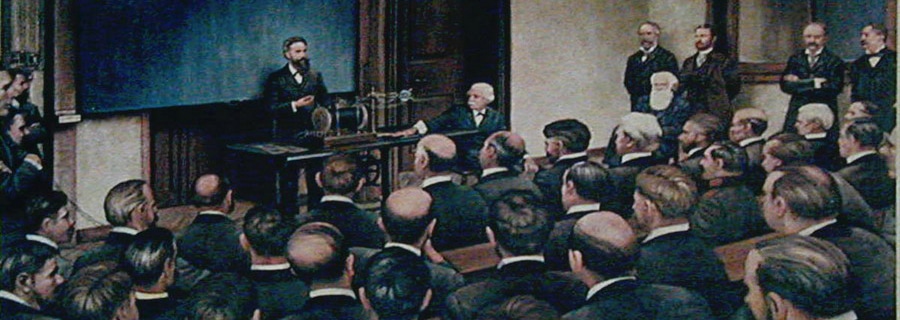
Wilhelm Conrad Röntgen’s first public presentation of the new X-rays took place on the 23rd of January 1896 in Würzburg.
The Swiss anatomist Albert von Kölliker volunteered to have his hand X-rayed during the lecture, and the resulting image has now been analysed with a new version of BoneXpert, presented in the Archives of Osteoporosis
The new method automatically measures the cortical thickness in the three middle metacarpals and was developed in cooperation with Jena and Copenhagen universities. Reference curves for four indices of cortical bone were determined for males and females up to the age of 90 based on a large cohort of healthy subjects from Jena.
The image of von Kölliker’s hand is now a classical specimen in the history of medical physics. We chose to celebrate the 120th anniversary of X-rays by analysing the image with the modern CAD software. It underlines that this imaging modality was established very quickly and has changed only little over time.

The metacarpal index of 78-year old Albert von Kölliker was found to be 0.634, which translates into a Z-score of 2.4. We conclude that his bone health was far above normal.
The clinical applications of the method have been documented extensively in studies based on the original implementation from 2001:
A) Osteoporosis screening: It has been shown that the method predicts hip fractures
B) Assessment of progression of rheumatoid arthritis
C) Monitoring of bone health in patients with growth hormone deficiency in the transition from childhood to adulthood
If you are interested in collaborating with us to develop this into a tool for clinical practice as part of the BoneXpert Server solution, or if you are interested in research projects, please contact Visiana.
——————————————————————————————————————————————————————-
On the technical side, the new method features several advantages over the original implementation:
1) A new and more powerful method is used to localise the metacarpals.
- The entire contour, rather than just the shafts, is reconstructed, which allows the measurement region to be placed relative to the ends of the bone and with a size scaled to the bone length. Hence, the measurement regions are anatomically consistent over the complete age span from infants to adults.
- The new method is more robust towards disturbances in image quality and abnormal bone structure – it so to speak understands the image better – and it automatically validates the measurement in more than 97% of the cases; only for the remaining 3% is an expert user needed to verify the result.
2) Reference curves for four indices of cortical bone are included – these are approximately defined as follows:
- The Metacarpal Index: MCI ∼ T / W. This works even if the image magnification is unknown as was the case for von Kölliker
- The Bone Health index: BHI ∼ T / (W L)0.333 This has the smallest relative SD for normal subjects of a given age
- The Volume Per Area: VPA ∼ T This is proportional to areal BMD
- The Exton Smith Index: ESI ∼ T / L
3) The method is implemented as a local server software, and the result is automatically stored in PACS. This gives a very easy workflow and it avoids sending the image away.