How well does Bone Health Index correlate with DXA?
The journal “Bone” brings a new paper entitled “Which skeletal imaging modality is best for assessing bone health in children and young adults compared to DXA? A systematic review and meta-analysis”, by Heba Shalof, Paul Dimitri, Farag Shuweihdi, and Amaka C Offiah.
The paper reviews the literature for the following three methods, and performs a meta-analysis of their correlation to DXA (dual-energy X-ray absorptiometry):
- QUS (Quantitative bone UltraSound), 17 paper
- pQCT (peripheral Quantitative Computed Tomography), 5 papers
- DXR (Digital X-ray Radiogrammetry), 8 papers
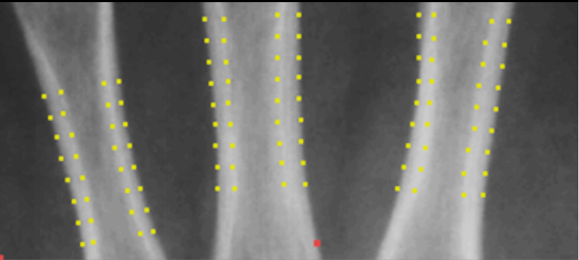
The DXR method is implemented in BoneXpert as the Bone Health Index, BHI.
The meta-analysis demonstrates that the strongest correlation is between DXR and DXA, with a coefficient of 0.71, and this leads the authors to the following statement:
“Of the imaging modalities compared to DXA in this review, DXR appears to have the most promise, being inexpensive, widely available, and can be used easily in children because it is a quick technique with software available that allows reliable and computerized measurement of BHI. Additionally, BHI describes not only cortical thickness (T), which represents aBMD, but also measures metacarpal length and width, which represent vBMD (non-size dependence method). Furthermore, DXR is considered a safe tool for bone health in children because its radiation dose (to a peripheral site) is relatively low. The ionizing radiation dose for a PA radiograph of the hand is <0.1 μSv [DXA radiation dose (1–6 μSv)].”